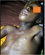
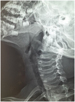
Cavum cysts, or nasopharyngeal cysts, are rare benign lesions, often of congenital origin, developing in the nasopharynx. Their discovery is frequently incidental during radiological or endoscopic examinations due to their asymptomatic nature. However, when they are large or poorly localized, they can cause symptoms such as nasal obstruction, rhinorrhea, recurrent serous otitis, or swallowing disorders. From an etiopathogenic perspective, they can result from the persistence of embryonic structures such as the Tornwaldt duct or from glandular obstruction. Diagnosis is primarily based on imaging, particularly MRI, and treatment is primarily surgical. This study reports a clinical case observed in the ENT department of Nianankoro Fomba Hospital in Ségou. This is a 7-year-old boy who has been suffering from inspiratory dyspnea, nasal obstruction, mouth breathing, postnasal drip, and nocturnal snoring for the past year. Clinical examination reveals good general condition but failure to thrive. Nasofibroscopy identifies a smooth, rounded, fluctuating mass partially occupying the cavum. Bilateral serous otitis media is also found on otoscopy. X-rays show a retronasal fluid-filled swelling. Endoscopic nasal excision is performed under general anesthesia. The postoperative course is favorable, with resolution of obstructive symptoms within the first few days. Histopathological analysis confirms a benign mucosal cyst. No signs of recurrence have been observed after six months of follow-up. This case illustrates the importance of endoscopic and radiological diagnosis in symptomatic forms of cavum cysts, and the good response to minimally invasive surgical treatment.
| Published in | International Journal of Otorhinolaryngology (Volume 11, Issue 2) |
| DOI | 10.11648/j.ijo.20251102.11 |
| Page(s) | 9-11 |
| Creative Commons |
This is an Open Access article, distributed under the terms of the Creative Commons Attribution 4.0 International License (http://creativecommons.org/licenses/by/4.0/), which permits unrestricted use, distribution and reproduction in any medium or format, provided the original work is properly cited. |
| Copyright |
Copyright © The Author(s), 2025. Published by Science Publishing Group |
Cavum Cyst, Snoring, Nasal Obstruction
ENT | Ear, Nose, and Throat |
MRI | Magnetic Resonance Imaging |
| [1] | Som PM, Curtin HD. Head and Neck Imaging. 5th ed. Elsevier; 2011. (chapter on cystic lesions of the nasopharynx). |
| [2] | Cure JK, Schmalfuss IM, Mancuso AA. “Tornwaldt cyst: MR imaging features." AJR Am J Roentgenol. 1996; 167(2): 481-484. |
| [3] | Alkan Z, Kiliç A, Akkaya H, Özlügedik S. " Nasopharyngeal retention cysts: Prevalence and clinical significance." Auris Nasus Larynx. 2015; 42(5): 396-399. |
| [4] | Kim KH, Sung MW, Kim YS, et al. "Clinical study of Tornwaldt's cyst." Acta Otolaryngol. 2002; 122(3): 320-324. |
| [5] | Dohar JE, Bonilla JA. Assessment and management of the child with nasal obstruction. Pediatr Clin North Am. 2003; 50(2): 359-372. |
| [6] | Harnsberger HR. Diagnostic Imaging: Head and Neck. 2nd ed. Elsevier; 2011. |
| [7] | Shugar JM, Som PM. Benign lesions of the nasopharynx in children: radiologic-pathologic correlation. Radiol Clin North Am. 1993; 31(4): 769-788. |
| [8] | Derkay CS, Darrow DH. Pediatric otolaryngology: airway and respiratory disorders. Pediatr Clin North Am. 2003; 50(2): 445-472. |
| [9] | Rupa V, Krishnaswami H, Job A. Endoscopic evaluation of nasopharyngeal masses in children. Int J Pediatr Otorhinolaryngol. 2002; 64(1): 59-66. |
| [10] | Eziyi JA, Ogunleye AO, Komolafe EO. Nasopharyngeal mucous retention cysts in children: case series and review of literature. Niger J Clin Pract. 2011; 14(3): 371-373. |
| [11] | Lee DH, Yoon TM, Lee JK, Lim SC. Surgical outcomes of endoscopic excision of benign nasopharyngeal masses. Braz J Otorhinolaryngol. 2016; 82(4): 398-403. |
APA Style
Haidara, A. W., Cissé, N., Coulibaly, D., Fofana, A., Sanogo, H., et al. (2025). A Cavum Cyst: A Case Report at the Nianakoro Fomba Hospital in Segou. International Journal of Otorhinolaryngology, 11(2), 9-11. https://doi.org/10.11648/j.ijo.20251102.11
ACS Style
Haidara, A. W.; Cissé, N.; Coulibaly, D.; Fofana, A.; Sanogo, H., et al. A Cavum Cyst: A Case Report at the Nianakoro Fomba Hospital in Segou. Int. J. Otorhinolaryngol. 2025, 11(2), 9-11. doi: 10.11648/j.ijo.20251102.11
@article{10.11648/j.ijo.20251102.11,
  author = {Abdoul Wahab Haidara and Naouma Cissé and Demba Coulibaly and Aminata Fofana and Harouna Sanogo and Ali Dembelé and Moussa Dembelé and Bagouma Traoré and Mariam Sangare and Mahamadou Doumbia and Boubacar Sanogo and Oumou Coulibaly and David Dackouo and Djibril Samake and Youssouf Sidibé and Sidiki Dao and Fatogoma Issa Kone and Boubacary Guindo and Siaka Soumaoro and Kadiatou Singare and Mohamed Amadou Keita},
  title = {A Cavum Cyst: A Case Report at the Nianakoro Fomba Hospital in Segou
},
  journal = {International Journal of Otorhinolaryngology},
  volume = {11},
  number = {2},
  pages = {9-11},
  doi = {10.11648/j.ijo.20251102.11},
  url = {https://doi.org/10.11648/j.ijo.20251102.11},
  eprint = {https://article.sciencepublishinggroup.com/pdf/10.11648.j.ijo.20251102.11},
  abstract = {Cavum cysts, or nasopharyngeal cysts, are rare benign lesions, often of congenital origin, developing in the nasopharynx. Their discovery is frequently incidental during radiological or endoscopic examinations due to their asymptomatic nature. However, when they are large or poorly localized, they can cause symptoms such as nasal obstruction, rhinorrhea, recurrent serous otitis, or swallowing disorders. From an etiopathogenic perspective, they can result from the persistence of embryonic structures such as the Tornwaldt duct or from glandular obstruction. Diagnosis is primarily based on imaging, particularly MRI, and treatment is primarily surgical. This study reports a clinical case observed in the ENT department of Nianankoro Fomba Hospital in Ségou. This is a 7-year-old boy who has been suffering from inspiratory dyspnea, nasal obstruction, mouth breathing, postnasal drip, and nocturnal snoring for the past year. Clinical examination reveals good general condition but failure to thrive. Nasofibroscopy identifies a smooth, rounded, fluctuating mass partially occupying the cavum. Bilateral serous otitis media is also found on otoscopy. X-rays show a retronasal fluid-filled swelling. Endoscopic nasal excision is performed under general anesthesia. The postoperative course is favorable, with resolution of obstructive symptoms within the first few days. Histopathological analysis confirms a benign mucosal cyst. No signs of recurrence have been observed after six months of follow-up. This case illustrates the importance of endoscopic and radiological diagnosis in symptomatic forms of cavum cysts, and the good response to minimally invasive surgical treatment.},
 year = {2025}
}
TY  - JOUR
T1  - A Cavum Cyst: A Case Report at the Nianakoro Fomba Hospital in Segou

AU  - Abdoul Wahab Haidara
AU  - Naouma Cissé
AU  - Demba Coulibaly
AU  - Aminata Fofana
AU  - Harouna Sanogo
AU  - Ali Dembelé
AU  - Moussa Dembelé
AU  - Bagouma Traoré
AU  - Mariam Sangare
AU  - Mahamadou Doumbia
AU  - Boubacar Sanogo
AU  - Oumou Coulibaly
AU  - David Dackouo
AU  - Djibril Samake
AU  - Youssouf Sidibé
AU  - Sidiki Dao
AU  - Fatogoma Issa Kone
AU  - Boubacary Guindo
AU  - Siaka Soumaoro
AU  - Kadiatou Singare
AU  - Mohamed Amadou Keita
Y1  - 2025/07/18
PY  - 2025
N1  - https://doi.org/10.11648/j.ijo.20251102.11
DO  - 10.11648/j.ijo.20251102.11
T2  - International Journal of Otorhinolaryngology
JF  - International Journal of Otorhinolaryngology
JO  - International Journal of Otorhinolaryngology
SP  - 9
EP  - 11
PB  - Science Publishing Group
SN  - 2472-2413
UR  - https://doi.org/10.11648/j.ijo.20251102.11
AB  - Cavum cysts, or nasopharyngeal cysts, are rare benign lesions, often of congenital origin, developing in the nasopharynx. Their discovery is frequently incidental during radiological or endoscopic examinations due to their asymptomatic nature. However, when they are large or poorly localized, they can cause symptoms such as nasal obstruction, rhinorrhea, recurrent serous otitis, or swallowing disorders. From an etiopathogenic perspective, they can result from the persistence of embryonic structures such as the Tornwaldt duct or from glandular obstruction. Diagnosis is primarily based on imaging, particularly MRI, and treatment is primarily surgical. This study reports a clinical case observed in the ENT department of Nianankoro Fomba Hospital in Ségou. This is a 7-year-old boy who has been suffering from inspiratory dyspnea, nasal obstruction, mouth breathing, postnasal drip, and nocturnal snoring for the past year. Clinical examination reveals good general condition but failure to thrive. Nasofibroscopy identifies a smooth, rounded, fluctuating mass partially occupying the cavum. Bilateral serous otitis media is also found on otoscopy. X-rays show a retronasal fluid-filled swelling. Endoscopic nasal excision is performed under general anesthesia. The postoperative course is favorable, with resolution of obstructive symptoms within the first few days. Histopathological analysis confirms a benign mucosal cyst. No signs of recurrence have been observed after six months of follow-up. This case illustrates the importance of endoscopic and radiological diagnosis in symptomatic forms of cavum cysts, and the good response to minimally invasive surgical treatment.
VL  - 11
IS  - 2
ER  -
ENT and Head and Neck Surgery Department, Nianankoro Fomba Hospital, Segou, Mali